


Services by Sunshine Neurology
Medical Services
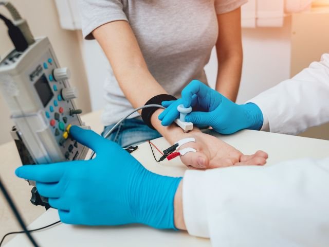
Electromyography (EMG)
Unlike other facility Dr.Patel himself performs EMG/ NCV test to provide highest quality and
accuracy.. EMG/ NCV test is used for diagnosis of various muscle and nerve disorders like
neuropathy, myopathy, carpal tunnel syndrome, radiculopathy, degenerative nerve diseases
such as amyotrophic lateral sclerosis (Lou Gehrig’s disease); and neuromuscular diseases (which
affect both nerves and muscles), such as myasthenia gravis.
Purpose of the EMG
- To aid in the diagnosis of primary muscle disorders, such as myopathy and muscular dystrophy; degenerative nerve diseases,, such as amyotrophic lateral sclerosis (Lou Gehrig’s disease); and neuromuscular diseases (which affect both nerves and muscles), such as myasthenia gravis
- To diagnose disorders that affect the neuromuscular junction (the connection between a nerve fiber and the muscle it supplies)
- To diagnose disorders of the nerves or nerve roots caused by nerve damage or ongoing nerve injury
- To detect blockages or slowed responses to nerve stimulation
- To detect the cause of pain, numbness, tingling, weakness or cramping in the arms or legs
Special Concerns about EMG
- People with a widespread skin infection should not take this test because the infection may pass into the muscle via the needle electrodes.
- Because the needles may cause intramuscular bleeding, people with bleeding disorders or those who take anticoagulants are advised caution while undergoing this procedure.
- Obesity or edema (swelling) may make it difficult to insert the electrodes into the muscle.
- A variety of medications may interfere with the results of this test.
Before the EMG - Do not use creams or lotions on the day of the test because it will interfere with the attachment of the electrode to the skin.
What You Experience
- You will either lie down on a bed or sit in a chair, depending on which muscles/nerves are being tested.
- The skin over the selected muscle is cleansed with an antiseptic, and a small needle electrode—which is very thin and resembles a stick pin—is inserted into the muscle. A reference electrode is inserted under the skin nearby. You may experience some discomfort due to the needles.
- The electrodes record the electrical activity while the muscle is relaxed. You will then be asked to contract the muscle gradually, with increasing forcefulness. The impulses are transmitted to a machine that amplifies the signals and displays them. Sometimes, the signals are converted into audio and played through a speaker.
- The procedure may be repeated at other locations.
- EMG generally takes about 25 to 30 minutes.
Risks and Complications of EMG
Rarely, blood may collect and clot under the skin (hematoma) at the needle insertion sites, causing swelling and discoloration. This is harmless but may cause some discomfort.
After the EMG
- You may be given pain-relieving medication to relieve any soreness around the electrode insertion sites.
- Resume your normal activities and any medications withheld before the test.
If a large hematoma develops at the needle insertion sites, apply ice initially; after 24 hours, use warm, moist compresses to help dissolve the clotted blood.
Results of EMG
- Your doctor will consider the test results—as well as your symptoms, your physical exam, and the results of other tests—in order to diagnose any muscle disease and identify the muscles affected.
- If a definitive diagnosis can be made, appropriate therapy will be initiated.

Electroencephalograph (EEG)
An electroencephalogram (EEG) is a test used to find problems related to the electrical activity of the brain. In electroencephalogram (EEG), small metal sensors (electrodes) are applied to your scalp to record the electrical signals produced by nerve cells in the brain. The electrodes transmit this information to a machine that magnifies the electrical activity and provides a graphic representation of brain waves.
In electroencephalogram (EEG), small metal sensors (electrodes) are applied to your scalp to record the electrical signals produced by nerve cells in the brain. The electrodes transmit this information to a machine that magnifies the electrical activity and provides a graphic representation of brain waves. This test is usually done while you are awake, but in one variation (called a sleep-deprived EEG), you must stay awake the night before, and brain activity is then recorded the next day.
Purpose of the Electroencephalogram
- To identify and evaluate the seizure focus and different types of seizure
- To evaluate brain wave activity in people with brain or spinal cord infections (such as meningitis or encephalitis), head injury, and psychiatric conditions
- To monitor brain wave activity during brain surgery.
- To help evaluate sleep disorders.
- To aid in the diagnosis of coma.
Who Performs Electroencephalogram
- A trained technician performs an EEG, and then the doctor reviews the test
Special Concerns about Electroencephalogram
- This test is usually conducted in a special room designed to eliminate electrical interference and minimize distractions.
- A variety of medications (including sedatives, anticonvulsants, tranquilizers, and barbiturates), caffeinated beverages, failure to eat before the test, or the presence of bright or flashing lights may alter the results of the test.
- You must remain still during the test. Any excess movement can interfere with the accuracy of the results.
Before the Electroencephalogram
- Inform your doctor of any medications you regularly take. Certain of these agents may need to be discontinued for 1 or 2 days before the test.
- Wash your hair the night before the test, and do not use hair spray, gel, or other hair care products after shampooing.
- Be sure to eat a normal meal before the test since low blood sugar (hypoglycemia) may interfere with test results. However, you should avoid caffeine-containing beverages, such as coffee, tea, or cola, for 8 hours before the test.
- You are asked to stay up as late as possible on the night before a sleep EEG
- If you are at risk for seizures, arrange for transportation to and from the test.
What You Experience during Electroencephalogram
- You will either sit in a reclining chair or lie down on a bed.
- For most studies, 15 to 20 electrodes are attached to your scalp with a special adhesive paste or gel in a specified pattern, plus one on each earlobe and one on the forehead.
- You are asked to relax, close your eyes, and lie still as the EEG recording is being taken. The examiner will note any movements, such as blinking, swallowing, or talking, that may affect the test results.
- Periodically, the examiner will stop the procedure to allow you to reposition yourself and get comfortable. Fatigue and restlessness can alter brain wave patterns.
- You may be asked to breathe deeply and rapidly (hyperventilation) for a few minutes to stimulate hidden brain wave abnormalities not evident during the resting EEG.
- “Photostimulation” may also be conducted by flashing a light at variable speeds over your face when your eyes are open or closed. This is intended to produce abnormal activity present in seizures that are stimulated by light.
- The procedure typically takes 45 to 60 minutes.
After the Electroencephalogram
- The electrodes are removed and the adhesive paste is washed away with acetone or warm water. You may need to use acetone and wash your hair at home to remove any residue.
- Unless you are actively having seizures or you underwent a sleep EEG, you may drive home immediately.
- Resume taking any medications withheld before the test, according to your doctor’s instructions.
Results of Electroencephalogram
- A neurologist will evaluate your brain wave patterns for evidence of any abnormality. For example, seizure activity is indicated by rapid, spiking waves, while cerebral lesions such as tumors or blood clots will result in abnormally slow EEG waves. The presence of an inflammatory brain disease, such as meningitis or encephalitis, may cause diffuse and slow brain waves.
- If a definitive diagnosis can be made, appropriate therapy will be initiated.
- In most cases, additional imaging tests of the brain—such as a CT scan, an MRI, or a nuclear scan—are necessary to pinpoint the location of any abnormality.
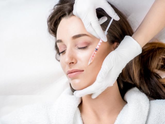
Botox
Botox, the best-known form of Botulinum toxin, has been increasingly used in Neurology practice since its first approval by FDA in 1989. In addition to its popular role for cosmetic purposes, it has been used in multiple neurological conditions, including migraine, tension headache, tilted neck (torticollis or cervical dystonia), chronic neck and back pain, facial and eyelid twitches (facial and blepharospasms); limb spasticity after stroke, cerebral palsy, multiple sclerosis or spinal cord lesions; hyperhidrosis; temple-mandibular joint dysfunction (TMJ) and tremors.
We offer botox treatment for following condition:
Cosmetic ( enahaced facial apperiance)
Migraine headache
Movement disorder

Calmare therapy
Calmare device is non‐invasive medical device for the treatment of high‐intensity oncologic and neuropathic pain, including pain resistant to morphine and other drugs. The device, with a biophysical rather than a biochemical approach, uses a multi-processor able to simultaneously treat multiple pain areas by applying surface electrodes to the skin. The device creates and sends a no-pain signal which becomes the dominant signal received by the brain, thus overriding the pain signal and providing pain relief for the patients.
Peripheral neuropathy (often referred to simply as “neuropathy“) refers to any condition that damages or disrupts nerves in the peripheral nervous system. Patient experience various symptoms like numbness, tingling, electric shooting pain, tightness, muscle cramps, restless leg. Most commonly in neuropathy there disruption of blood supply to nerve leading nerve degeneration on nerve over time.
Calmare device is non‐invasive medical device for the treatment of high‐intensity oncologic and neuropathic pain, including pain resistant to morphine and other drugs. The device, with a biophysical rather than a biochemical approach, uses a multi-processor able to simultaneously treat multiple pain areas by applying surface electrodes to the skin. The device creates and sends a no-pain signal which becomes the dominant signal received by the brain, thus overriding the pain signal and providing pain relief for the patients.
Treatment using the Calmare device avoids the harmful, potentially fatal, adverse side effects and addictive properties linked to narcotic pain killers.
The Calmare device has been used to successfully treat over 4,000 patients worldwide, where it has been shown to be effective in treating neuropathic and oncologic pain. Conditions treated include:
- Chemotherapy-induced peripheral neuropathy
- Phantom limb syndrome
- Sciatica
- Post-surgical neuropathic pain
- Low back pain
- Neck pain
- Reflex sympathetic dystrophy
- Post herpetic neuralgia (PHN)
- Peripheral Neuropathy
Calmare therapy is major innovation in pain treatment and recently mayo clinic also started using this device. Calmare therapy currently offers by few locations in US. We are pleased to offer this unique treatment for neuropathy at Sunshine Neurology by Dr. Kamlesh Patel, MD.
You can find out more about calmare therapy and watch video at calmarett.com and calmarerelief.com
Neurological Exam
ALS/Lou Gehrig’s disease | |
| Alzheimer’s disease | Multiple sclerosis (MS) |
| Back pain | Muscle disease
Motor Neuron Disease |
| Bell’s palsy | Nerve injuries/numbness |
| Carpal tunnel syndrome | Peripheral Neuropathy |
| Chronic pain | Parkinson’s disease |
| Dementia | Spinal cord injury |
| Dizziness | |
| Epilepsy (seizure) | |
| Headaches/migraines | Strokes/TIA |
| Hemifacial spasm | Tics/Tourette’s syndrome |
| Memory Impairment | Tremor |
| Movement disorders | Vertigo |


Neurocognitive Assessment
The brain is complex. It monitors the state of the body and its environment, controls the body’s motion, and also performs many of the higher-level cognitive functions that allow us to think, plan, and accomplish the tasks in our daily lives. Cognitive functions are the basis for our activities of daily living and include the ability to shop, talk on the telephone, manage finances, drive, and learn something new. While it is true that most brain functions involve much coordination between different brain regions, we also know that certain brain functions can be tested separately. Neuropsychologists have defined a set of essential cognitive functions that underlie brain wellness.
• Memory
• Executive Function
• Attention
• Visual Spatial
• Verbal Function
• Problem Solving
• Working Memory
In addition, BrainCare offers questionnaire-based scales for psychosocial factors that may impact brain wellness.
What is the client’s experience like?
BrainCare testing is very user-friendly and requires little orientation. Although the testing is performed on a computer, it does not require the client to know how to use one. Testing times typically range from 45-60 minutes, depending on the patient’s age.
How exactly does BrainCare measure brain wellness?
BrainCare assesses the client’s brain wellness by precisely measuring the patient’s performance on a series of interactive tests – measuring both accuracy and response time. Test results are compared to previous data from the same patient and performance data from that of a “normative” peer group (i.e., age and education appropriate). The result is a profile of cognitive areas that are ‘strong’ and those that are relatively ‘weak’. The relatively weak areas are the ones that should be targets for exercises that can be done at home.
With BrainCare, patients and their families have access to reports on patient test performance, as well as activity recommendations based on the latest clinical research. Clinicians can more easily provide research-backed recommendations to help patients better track progress.
Proven Science
Research involving tests by NeuroTrax, the company behind BrainCare, has been published in over 75 peer-reviewed scientific articles. In all, over 190 research studies have been performed involving over 28,000 subjects from 16 countries. Also, over 100 international conference presentations have featured our tests. No other brain fitness testing system has as much scientific backing.
Precision
BrainCare is based on optimized technology that achieves accurate measurements of response times. That translates to better estimates of how quickly the brain can process and analyze information – a key dimension of brain fitness.
Track Performance
BrainCare is easy to re-administer, making it ideal for tracking longitudinal changes. In addition, there are features to minimize practice effects.
Practicality
We designed BrainCare tests to work in the “real world” and ensure that accurate data are gathered as long as the testing computer fits the basic technical requirements. For example, our system conducts practice sessions before each test and automatically determines whether a client has adequately mastered the “mouse” buttons and has the visual acuity and color recognition necessary to perform the tests.
No prior computer experience is necessary
As reported in a study involving nearly 3,000 older people, most clients report that our tests are easy to use. This was true even for people with no computer experience.